USG of Gall Bladder
Protocols
🌏Normal Gall Bladder Usg findings :
- Length : 12 cm
- Width: 4 cm
- Wall Thickness : less then 3 mm

🌏কখন usg তে gall bladder দেখা যাবে না ?
1.Post Cholecystectomy state
2.Physiological contraction (after meal.as gall bladder
becomes empty after meal specially after fatty meal)
Acute cholecystitis Usg findings :


1. Gall bladder wall thickened. Wall thickness >3 cm
2. Hypoechoic wall (কালো হবে)
3. Stone or sludge may be present.
4. Pericholecystic fluid may be present.
Chronic cholecystitis Usg findings :

1. Wall thickened.
2.Gall bladder may be small.
3.Hyperechoic wall (due to fibrosis)
4. Stone may be present.
USG findings of Empyema:

Gall bladder :
1.Sloughing of mucosa of gall bladder
2. Gal bladder is filled with debris
Cholelithiasis USg findings :
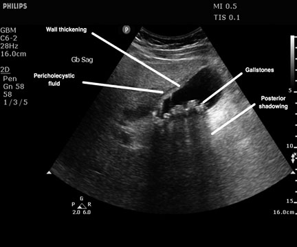
1.Bright echogenic structure
2.posterior aquestic shadow.
3.Moves with change of posture if not impacted at neck of gall bladder
🌏Billiary/gall bladder Sludged usg findings :

1.Echogenic structure
2. No posterior aquestic shadow
3.It has linear wall
4. Some times echiogenicity may be isoechoic to liver.
5. Moves with posture change.
🌏Usg Findings of Mucocele gall bladder:

1.Length increase >10 cm
2. Gall bladder wall regular
3. Width increase > 4 cm
4. Contains clear fluid