USG of Liver
Protocols
Normal Liver measurement of Usg:

Right Lobe of Liver :
1.Mid-clavicular line থেকে length 15 cm. এই length বয়স অনুসারে ভিন্ন হয়।
এই liver lenght বেড়ে গেলে আমারা hepatomegally বলি।
2.Anterior -posterior Diameter: 10 cm

Left lobe of liver:
1.Midline বরাবর এই length 10 cm.
2.Anterior -posterior Diameter: 6 cm
Hepatic Vein USG তে পাতলা বা thin walled দেখাবে।
portal Vein Usg তে মোটা or thick walled দেখাবে।
🛑কোন Technique Liver usg করবো।
A.যদি Sagital or longitudinal scan করি ঃ
 তাহলে আমরা,
1.Right lobe এর ক্ষেত্রে mid clavicular line বরাবর দেখবো।
2.Left lobe এর ক্ষেত্রে mid line বরাবর measure করবো।
B.যদি Coronal/Transverse plan usg করিঃ

তাহলে আমরা
🖍️Right lobe & Left lobeAnterior -Posterior Diameter দেখবো।
Liver Pathology in USG COLOR:
🌏Liver Abscess এর usg findings :
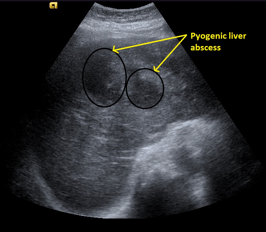
- circumscribed area(গোলাকার, পাশের ছবিতে দেখেন
- Margin: regular / indetermined(বেশির ভাগ ক্ষেত্রে নাও থাকতে পারে)
- Posterior enhancement -থাকে না
- Echogenic ( white) - if gas forming organish দ্বারা liver abscess হয়
- Doppler- ভিতরে কোন blood flow দেখা যায় না।


Fatty Liver Usg Findings :পাশের ছবিতে লক্ষ্য করুন।
- Steatosis/fatty liver manifests as increased echogenicity.
- renal cortex appearing relatively hypoechoic compared to the liver parenchyma (normally liver and renal cortex are of a similar echogenicity)
- increased echogenicity relative to the spleen, when there is parenchymal renal disease
- absence of the normal echogenic walls of the portal veins and hepatic veins
- important not to assess vessels running perpendicular to the beam, as these produce direct reflection and can appear echogenic even in a fatty liver
- poor visualization of deep portions of the liver
- poor visualization of the diaphragm
🌏Fatty liver Grading:
grade I: Increased hepatic echogenicity with visible periportal and
diaphragmatic echogenicity
grade II: Increased hepatic echogenicity with imperceptible periportal
echogenicity, without obscruation of diaphragm
grade III: Increased hepatic echogenicity with imperceptible periportal
echogenicity and obscuration of diaphragm
🌏Acute Hepatitis usg Findings :
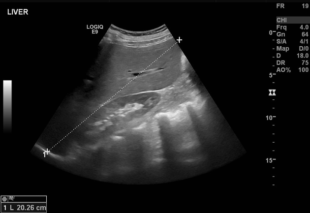
1.hepatomegaly (most sensitive sign) >15.5 cm at the midclavicular line 4
2.starry sky appearance has been found to have poor sensitivity and specificity
3.gallbladder wall thickening variably present may be more closely associated with hepatitis
3.periportal edema accentuated brightness of portal vein radicle walls
4.color/spectral Doppler: normal the overall echotexture is often decreased.
🌏Liver Cirrhosis Usg findings :

1.overall coarse and heterogeneous echotexture
2.segmental hypertrophy/atrophy (পাশের ছবিতে দেখুন)
3.caudate width: right lobe width >0.65
4.reduction of the transverse diameter

🛑signs of portal hypertension
🕹️Doppler flow changes
⛔portal venous system
1.enlarged portal vein: >13 mm
2.slow portal venous flow
3.reversal or to-and-fro portal venous flow
4. portal venous thrombosis +/- cavernous transformation
⛔splenomegaly
⛔ascites
⛔fatty change (variable)
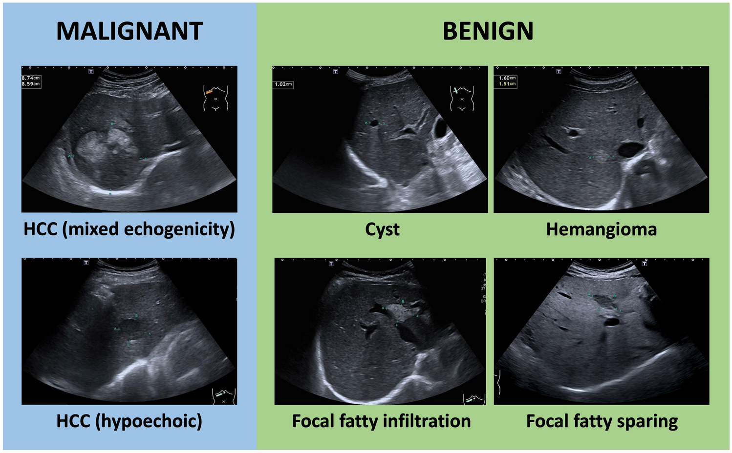
🌏HCC Usg findings :
🕹️small focal HCC appears hypoechoic compared with normal liver
🕹️larger lesions are heterogeneous due to fibrosis, fatty change, necrosis and calcification
🕹️a peripheral halo of hypoechogenicity may be seen with focal fatty sparing
🕹️diffuse HCC may be difficult to identify or distinguish from background cirrhosis

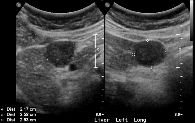
🌏Hepatic Cyst USg findings :

Fig-1: Hepatic cyst(Hydatid cyst)

Fig-2: Hepatic cyst
1. Number: 1 / cluster of cyst
2. Shape: rounded / oval
3. Margin: well defined, smooth, thin walled
4.Anechoic inside & show edge shadow
5.Through transmission (posterior enhancement)
6. If infected or haemorrhagic show internal echos or septation.
7.Size