Electroencephalography(EEG)
Protocols
EEG Definition:
EEG stands for “electroencephalography” which is an electrophysiological process torecord the electrical activity of the brain. EEG measures changes in the electrical activityproduced by the brain. Voltage changes come from ionic current within and between some brain cells called neurons.

What is an EEG?
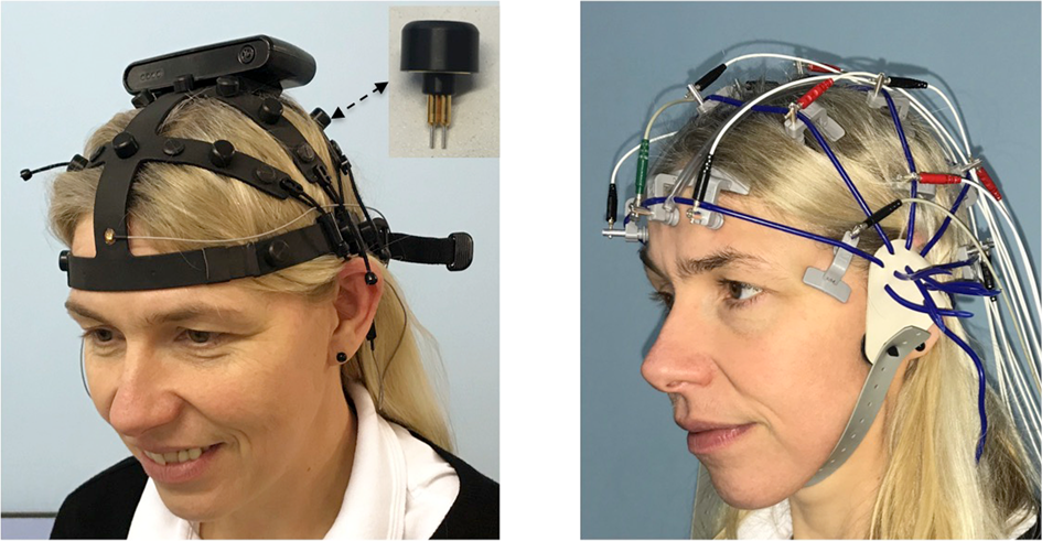
An EEG test evaluates the electrical activity of the brain. EEG scans are performed by placing EEG sensors —
small metal discs also called EEG electrodes — on your scalp. These electrodes pick up and record the
electrical activity in your brain. The collected EEG signals are amplified, digitized, and then sent to a computer
or mobile device for storage and data processing.
Analyzing EEG data is an exceptional way to study cognitive processes. It can help doctors
establish a medical diagnosis, researchers understand the brain processes that underlie human
behavior, and individuals to improve their productivity and wellness.
Types of Brainwaves that EEG Measures
The electrodes of an EEG device capture electrical activity expressed in various EEG frequencies. Using an algorithm called a Fast Fourier Transform (FFT), these raw EEG signals can be identified as distinct waves with different frequencies. Frequency, which refers to the speed of the electrical oscillations, is measured in cycles per second — one Hertz (Hz) is equal to one cycle per second. Brainwaves are categorized by frequency into four main types: Beta, Alpha, Theta and Delta.
The following paragraphs discuss some of the functions associated with the four main brain frequencies. These functions have simply been found to be associated with different brain frequencies — there is no one-to-one linear correspondence between a frequency band and a given function of the brain.

Beta Waves (frequency range from 14 Hz to about 30 Hz)
Beta waves are most closely associated with being conscious or in an awake, attentive and alert state. Low-amplitude beta waves are associated with active concentration, or with a busy or anxious state of mind. Beta waves are also associated with motor decisions (suppression of movement and sensory feedback of motion). When measured by an EEG device, the signals are often referred to as EEG beta waves.
Alpha Waves (frequency range from 7 Hz to 13 Hz)
Alpha waves are often associated with a relaxed, calm and lucid state of mind. Alpha waves can be found in the occipital and posterior regions of the brain. Alpha waves can be induced by closing one’s eyes and relaxing, and they are rarely present during intense cognitive processes like thinking, mental calculus and problem-solving. In most adults, alpha waves range in frequency from 9 to 11 Hz. When measured by an EEG device, these are often referred to as EEG alpha waves.
Theta Waves (frequency range from 4 Hz to 7 Hz)
Brain activity within a frequency range comprised between 4 and 7 Hz is referred to as Theta activity. Theta rhythm detected in EEG measurement is often found in young adults, particularly over the temporal regions and during hyperventilation. In older individuals, theta activity with an amplitude greater than about 30 millivolts (mV) is seen less commonly, except during drowsiness. When measured by an EEG device, these are often referred to as EEG theta waves.
Delta Waves (frequency range up to 4 Hz)
Delta activity is predominantly found in infants. Delta waves are associated with deep stages of sleep in older subjects. Delta waves have been documented interictally (between seizures) in patients with absence seizures, which involve brief, sudden lapses in attention.
Delta waves are characterized by low-frequency (about 3 Hz), high amplitude waves. Delta rhythms can be present during wakefulness — they are responsive to eye-opening and may be enhanced by hyperventilation as well. When measured by an EEG device, these are often referred to as EEG delta waves.
Different Types of EEG Devices
EEG machines come in the form of a few different wearable EEG devices. At the highest level is the
difference between clinical EEG devices (used in a healthcare and scientific research setting) and
consumer EEG devices (used in consumer research, academic research and performance and wellness).
With clinical devices, participants cannot move while wearing the device, and the data needs to be collected in
a controlled and shielded environment to avoid distorting the signal. Consumer EEG devices like EMOTIV’s
wireless headsets allow users to monitor brain activity anywhere.
The variation between different types of wearable EEG devices is necessary to support the requirements
of professionals who use EEG systems and the settings in which data is collected. For example,
neurologists and neuroscientists often need a higher density of sensors to perform their data analysis than
a consumer researcher might. In addition to EEG electrode placement, there are a few other notable
variations between EEG systems to consider.
EEG Caps VS. EEG Headsets
What’s the difference between an EEG cap and an EEG headset? The main difference between these two
most common types of wearable EEG devices is in the number of electrodes. Headsets usually range from
5-20 electrodes. Caps can support more sensors, as they have a larger surface area for electrode placement.
Advantages of EEG
Despite relatively low spatial sensitivity of EEG, it claims multiple advantages over some of the previously listed brain imaging and brain research techniques:
- EEG has a very high temporal resolution compared to fMRI. It can pick up the rapid reactions of the brain that happen at the speed of milliseconds, which allows it to sync accurately what happens in the brain and in the environment. EEG is recorded at sampling rates between 250 and 2000 Hz in clinical and research settings. More modern EEG data collection systems can record at sampling rates above 20,000 Hz if desired.
- Significantly lower hardware costs and total cost of ownership (TCO).
- EEG data is collected non-invasively unlike electrocorticography, which requires neurosurgery for electrodes to be placed directly on the surface of the brain.
- Mobile EEG sensors can be used in more places than fMRI, SPECT, PET, MRS, or MEG, as these techniques rely on heavy, costly and immobile equipment.
- EEG is silent, to allow for studying the responses to auditory stimuli.
- Compared to fMRI and MRI, there is no physical danger around an EEG machine. fMRI and MRI are powerful magnets that prevent use by patients with metallic gear such as pacemakers.
- fMRI, PET, MRS, and SPECT can aggravate claustrophobia which can corrupt test results. EEG does not induce claustrophobia as subjects are not confined to a small space.
- Consumer EEG scans allow for more subject movement during testing, unlike most other neuroimaging techniques.
- EEG does not involve exposure to radioligands, unlike positron emission tomography, or high-level magnetic fields like MRI or fMRI.
- EEG does not involve exposure to high-intensity (>1 tesla) magnetic fields.
- Compared to behavioral testing methods, EEG can detect covert processing (processing that does not require a response). This technology also is used in subjects who are unable to make a motor response.
- EEG has a low barrier of entry for consumer use so it is a powerful tool for tracking and recording brain activity during different activities of daily life, allowing for a nearly limitless number of applications.
- EEG sleep analysis can indicate significant aspects of the timing of brain development, including evaluating adolescent brain maturation.
- There is a better understanding of exactly what signal is measured with EEG, compared to the BOLD (Blood-oxygen-level-dependent) imaging used in fMRI.
EEG Use Cases
There are many modern applications for EEG measurement.
- Neuroscience
- Brain education programs
- Neuromarketing
- Sleep Studies
- Brain Computer Interface (BCI)
- Cognitive Performance
- Self-Quantification
- Emotional States
- ADHD Therapy
- Neurological disorders
- Brainwave entrainment
- Cognitive behavioral therapy
- Neuroinformatics
- Brainwave gaming
- AR & VR supplement
- Dysphagia and Dementia
- Stroke rehabilitation
- Working memory tests (N-back)