CT Scan
Protocols
CT Scan OF Intracerebral haematoma:

Plate 1A
Multiaxial CT scan of brain showing:
Irregular hyperdense lesion in left parietal region.
Perilesional
oedema.
Compression of left lateral ventricle.
Midline
shift to right.
Diagnosis: Intracerebral haematoma.
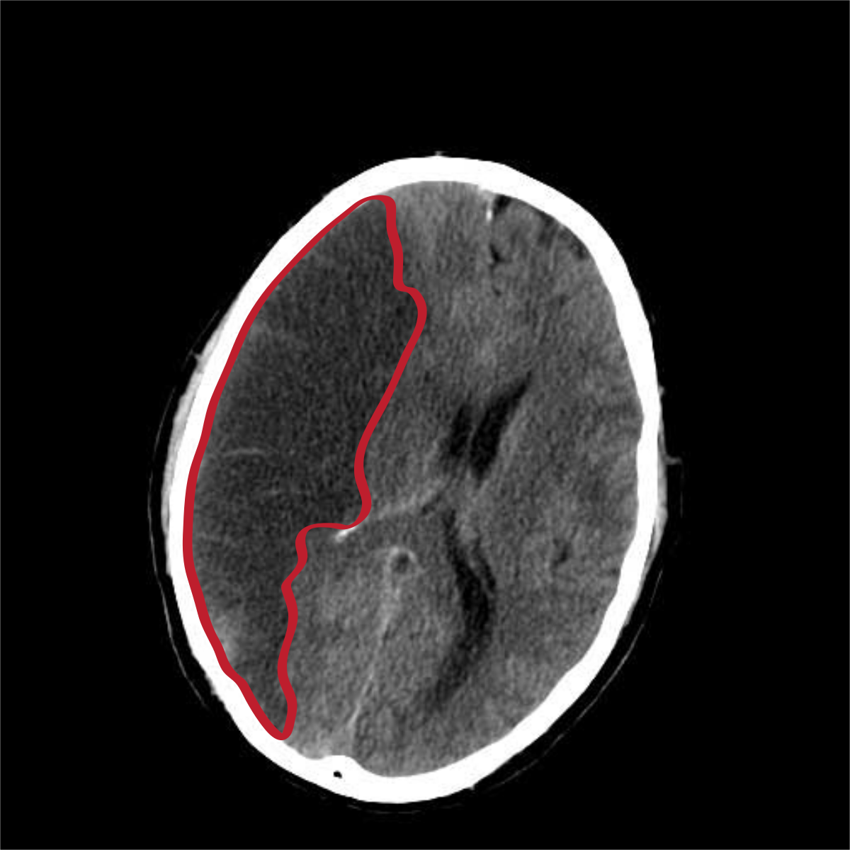
CT Scan of subarachnoid haemorrhage:


Findings:
CT scan of the head shows high attenuation (white
areas)
with obliteration of the ventricles. The diagnosis
is subarachnoid haemorrhage.
CT Scan of subdural hematoma :
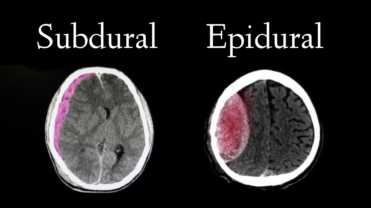
CT scan of the head shows crescent-shaped hyperdense
extra-cerebral collection within the subdural space. The
diagnosis is subdural hematoma on the left /Right side.

CT Scan of extradural haematoma:
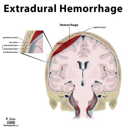

🛑CT scan of the head shows high density bi-convex area.
The diagnosis is extradural haematoma on the left/right side.
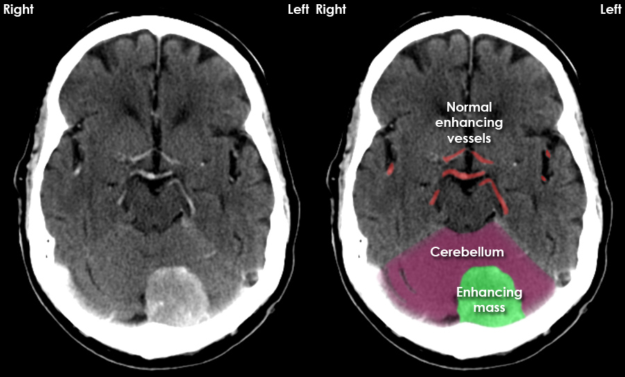
CT Scan of cerebral abscess:

Contrast CT scan of head showing a ring-like cystic
hypodense lesion on the right parietal lobe. The diag.
nosis is cerebral abscess.

CT Scan of hydrocephalus:
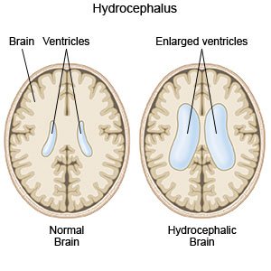

🛑CT scan of the head shows marked dilatation of both
lateral ventricles with thinned brain tissue outside the
hypodense area. The diagnosis is hydrocephalus.
CT Scan of cerebral infarction:

🛑CT scan of the brain shows hypodense area on the right
side with shifting of the ventricle to the left side. The
diagnosis is right-sided cerebral infarction.
CT Scan of meningioma:
🛑CT scan of the head shows hyperdense, irregular mass
in the frontal region. The diagnosis is meningioma.
CT Scan of liver abscess:
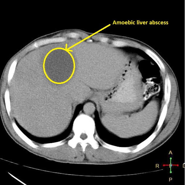
🛑CT scan of the abdomen shows multiple hypodense
area with ragged irregular margin within the right lobe
of the liver. The diagnosis is multiple liver abscess.
